
What Does an MRI Show for Cauda Equina Syndrome?
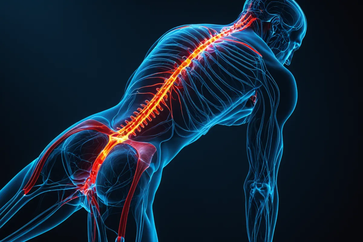
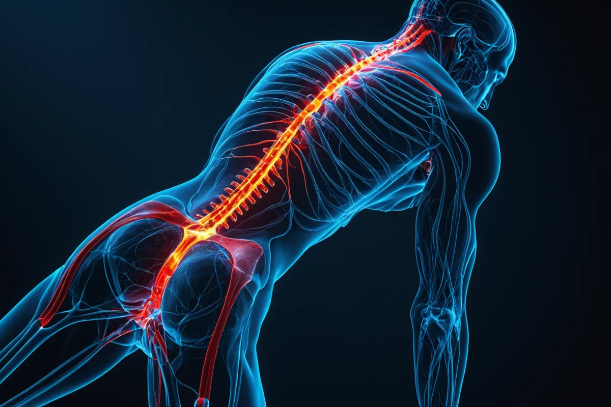
A cauda equina syndrome (CES) MRI is used to determine if spinal nerves are actually compressed, how severe the narrowing is, and whether surgery is urgent. Unlike simple disc issues causing only leg numbness, CES requires immediate attention when symptoms include urinary problems or loss of sensation around the anus.
Time is critical with CES; it’s better to confirm nerve compression on MRI rather than dismiss symptoms. However, a single MRI scan isn’t enough for diagnosis—it must align with symptoms, physical examination findings, and changes in neurological function.
What Nerve Compression Does a CES MRI Reveal?
The first thing doctors look at during a CES MRI is how narrow the spinal canal has become. When nerve bundles in the lower spine are compressed, this appears on both cross-sectional and sagittal views.
Doctors also check for common causes like large disc herniations, severe stenosis, hematomas, tumors, or inflammation. It’s not just about back pain—it’s whether the pathway nerves travel through is blocked.
For example, if a large disc protrudes backward at L4-5 or L5-S1, nearly filling the spinal canal, it raises stronger suspicion of CES rather than simple leg pain. In such cases, MRI reports often mention nerve compression, severe stenosis, or massive disc herniation.
Find more articles on similar topics in one place.
View Related ArticlesWhy CES Symptoms and MRI Must Be Evaluated Together
CES symptoms are more serious warning signs than simple back pain. Key indicators include numbness or weakness in both legs, reduced sensation around the saddle area, difficulty urinating, or involuntary urine leakage.
While MRI is important, relying solely on imaging can be misleading. Some people show changes on MRI but have mild symptoms, while others with ambiguous scans may experience rapid neurological decline. Diagnosis requires combining symptom assessment, physical exams, and MRI results.
For instance, if urination becomes difficult or bowel control changes, don’t dismiss it as a simple disc issue—seek prompt medical evaluation.
How MRI Shows the Cause of Cauda Equina Syndrome
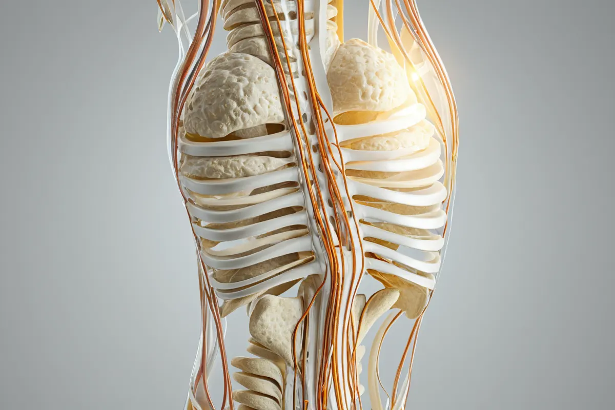
The causes of CES aren’t limited to one condition. The most common cause is a large disc herniation, but severe degenerative stenosis, spinal canal bleeding, tumors, or infectious inflammation can also be responsible.
MRI findings vary depending on the cause. Disc herniations show tissue pressing directly on nerves, stenosis appears as a narrowed passage, and hematomas look like acute masses.
For example, after a fall that causes sudden leg weakness and urinary issues, doctors must consider hematoma in addition to disc problems. Delaying diagnosis can limit treatment options.
Pitfalls in CES Diagnosis
Self-diagnosing CES based on online checklists isn’t reliable. Many people confuse common symptoms like constipation with CES, but true CES involves multiple signs: loss of sensation around the anus, urinary issues, and bilateral leg symptoms.
Constipation alone doesn’t indicate CES. However, if it’s accompanied by reduced saddle area sensation or urinary changes, further evaluation is needed. Conversely, even without severe back pain, diminished saddle sensation or weak urine stream should raise concern—walking ability doesn’t rule out nerve damage requiring MRI.
How MRI Informs Treatment Decisions
Treatment for CES depends on the severity of compression and its cause as seen on MRI. If a disc is causing pressure, emergency surgery may be considered; for infections or tumors, different approaches are needed.
The key questions doctors ask during MRI review are: Are nerves still functional? Is prompt decompression necessary? This makes symptom onset timing more critical than test results alone.
While some patients wait days for MRI results, those with urinary issues or leg weakness shouldn’t delay—seek urgent care at an emergency room, neurosurgery, or orthopedics clinic.
Monitoring Recovery After CES Treatment
After the acute phase, rehabilitation may follow. Even after surgery, some patients experience lingering sensory loss, muscle weakness, or urinary issues, with recovery varying widely between individuals.
MRI isn’t used to guide rehab but helps detect new compression or recurrence. If back pain returns along with worsening leg symptoms, don’t assume it’s just muscular—re-evaluation is needed.
For example, if walking is possible after surgery but foot sensation remains dull and urination feels different, report these changes immediately. They could signal a normal recovery phase or an issue requiring intervention.
When to Seek Immediate Medical Attention
If you experience any of the following, consider CES and seek prompt medical care:
- Sudden numbness or weakness in both legs
- Dull sensation around the saddle area
- Changes in urination (difficulty starting or leakage)
- Altered bowel control compared to usual
- Sudden loss of leg strength while walking
These symptoms shouldn’t be dismissed as simple back pain. MRI is a key diagnostic tool, but delaying care for results can worsen outcomes—prompt medical evaluation takes priority.
Misconceptions About CES MRI Interpretation
A common misconception is that terms like “stenosis” or “disc herniation” on an MRI report automatically mean CES. In reality, imaging findings must match symptom severity for a diagnosis.
Conversely, if the MRI appears mild but symptoms are classic, more aggressive evaluation is needed. Reports serve as reference—not final decisions—so clinical judgment and neurological exams guide treatment.
When researching online, focus on whether your symptoms signal an emergency rather than what specific findings appear on MRI. If urinary issues arise, act quickly.
FAQ
Q1. Does a CES MRI always require contrast?
Most cases can be assessed with standard lumbar MRI without contrast. However, for conditions like tumors, infections, or post-surgical evaluation, contrast may be added if needed.
Q2. Can CES symptoms exist even if the MRI appears normal?
Yes, this is possible. Early-stage symptoms or issues elsewhere might not show on MRI, so diagnosis requires combining results with physical exams and symptom progression.
Q3. Should constipation alone raise concern for CES?
No—constipation alone isn’t enough to suspect CES. Look instead for accompanying changes like saddle area numbness, urinary issues, or bilateral leg symptoms.
CES is a time-sensitive condition where early action matters most. If symptoms are suspected, don’t delay seeking medical advice. Diagnosis and treatment may vary based on current clinical guidelines, so consult your healthcare provider immediately if you experience any concerning symptoms.
Explore more articles on similar topics.
View Category ArticlesFAQ
Does a CES MRI always require contrast?
Most cases can be assessed with standard lumbar MRI without contrast. However, for conditions like tumors, infections, or post-surgical evaluation, contrast may be added if needed.
Can CES symptoms exist even if the MRI appears normal?
Yes, this is possible. Early-stage symptoms or issues elsewhere might not show on MRI, so diagnosis requires combining results with physical exams and symptom progression.
Should constipation alone raise concern for CES?
No—constipation alone isn’t enough to suspect CES. Look instead for accompanying changes like saddle area numbness, urinary issues, or bilateral leg symptoms.
Korean Source
This article is an English localization of a Korean-language report from BlogScore. Original: 마미증후군 MRI에서 무엇을 보나